

Custom HIFU Transducer Design for Enhanced Therapeutic Precision
2026-08-17
High-intensity focused ultrasound (HIFU) promises noninvasive therapy, yet most systems still rely on off-the-shelf transducers that force clinicians to compromise on beam shape, focal depth, and acoustic power. That compromise quietly undermines treatment precision—until now. Custom HIFU transducer design is rewriting the rules, and Siansonic is at the center of that shift. Instead of adapting treatment plans to hardware limitations, engineers can now match piezoelectric geometry, lens curvature, and array configuration to specific anatomical targets. The result? Sharper focus, tighter margins, and fewer unintended tissue effects. In this post, we explore how tailored transducer architectures are moving therapeutic ultrasound from a generalized tool to a patient-specific instrument—and why that matters for the future of noninvasive medicine.
Rethinking Focal Geometry for Patient-Specific Energy Delivery
Standard focal geometries in energy delivery have long been treated as fixed shapes—ellipses, cylinders, or spheres—that are simply scaled to fit a generic treatment volume. Yet real patient anatomy rarely conforms to such idealized forms. Rethinking focal geometry begins with mapping the irregular boundaries of a target lesion against surrounding healthy tissue, then designing an energy pattern that mirrors those contours rather than forcing the target into a predetermined mold.
This shift demands more than static contour matching. Tissue heterogeneity, respiratory motion, and organ deformation require focal geometry to behave as a living parameter that can be recomputed mid-treatment. Combining patient-specific imaging with fast optimization algorithms allows the focal zone to be reshaped slice by slice, concentrating energy where it is needed while creating steep gradients at critical structures that were previously difficult to protect.
Implementing such geometries in routine practice means reworking the entire planning chain, from dose calculation engines to delivery hardware calibration. Instead of relying on a library of pre-approved focal shapes, clinicians gain the ability to define energy fields that are as unique as the patient receiving them, reducing collateral damage and improving local control without lengthening treatment times.
Matching Acoustic Impedance to Tissue Heterogeneity Without Losing Focus
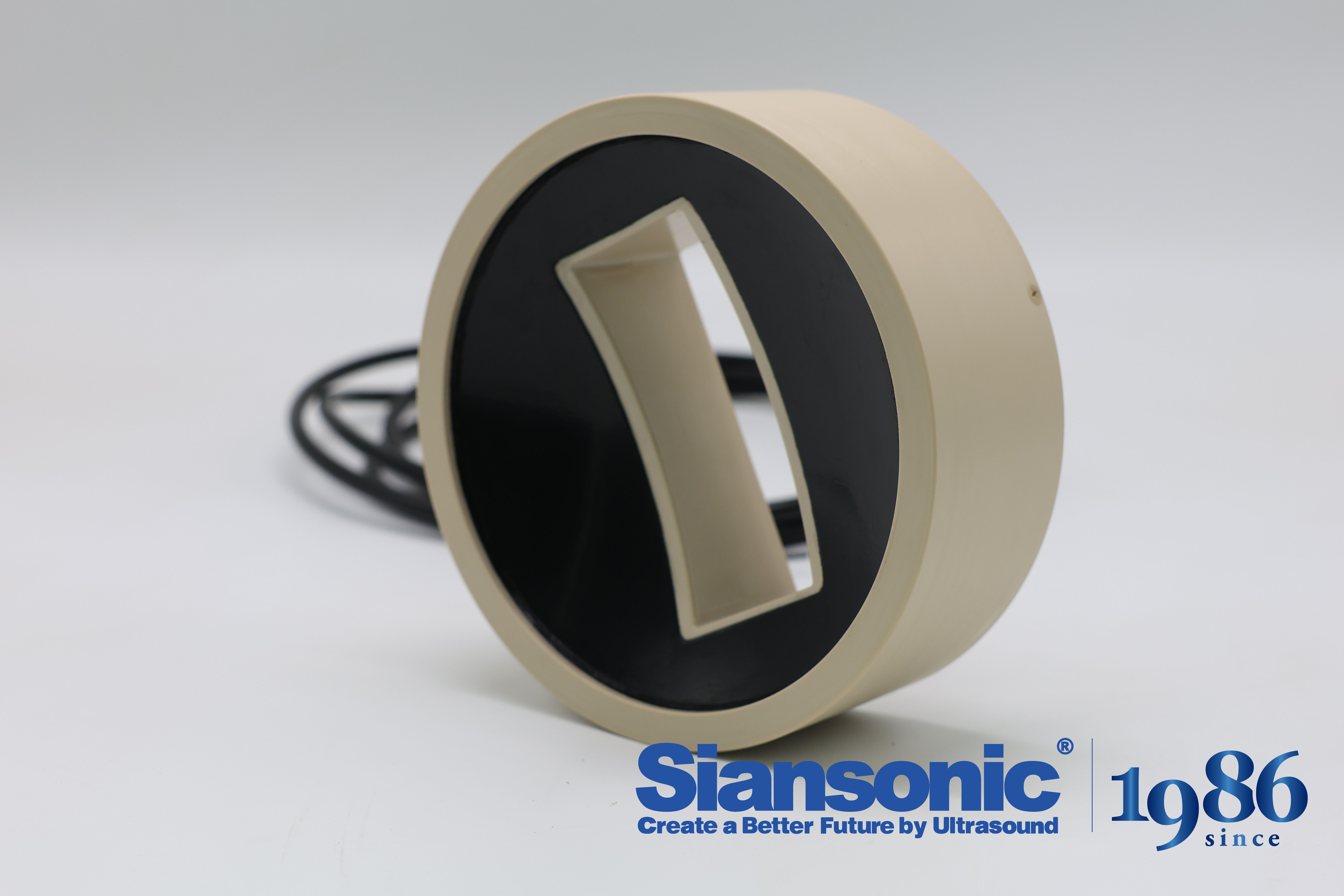
Tissue is never the homogeneous slab that simplified models assume. Fat, muscle, connective tissue, and fluid pockets each carry a different acoustic impedance, and when a focused beam crosses these boundaries, part of the wave reflects and the rest bends slightly off its intended path. The result is a focus that smears, shifts, or loses peak intensity. Matching impedance in this context is not about finding one magic value, but about accepting that the propagation path is a series of mismatched segments and designing the transducer or coupling layer to anticipate that variability.
One practical route is to replace a single rigid matching layer with a gradient or composite structure that transitions from the piezoelectric element to the skin without a sharp impedance jump. Thin layers of epoxy loaded with different volume fractions of metal oxide powders can create a staircase of acoustic impedances. Each step reflects less energy than a direct interface, but the real advantage is that a broader range of tissue impedances falls within the tolerable mismatch. This keeps the beam from losing too much energy at the surface while still allowing the deeper focus to remain tight, because the wavefront leaving the transducer is less distorted in the first place.
When the target sits behind ribs, scar tissue, or fatty regions, passive matching alone rarely restores a crisp focus. Here the array geometry and electronic phase correction become part of the impedance strategy. By measuring arrival time differences from a known scatterer or using a model-based estimate of the tissue inhomogeneity, the system can pre-distort the transmitted wave so that after passing through the irregular layers, the wavefront converges where intended. That does not eliminate impedance mismatch, but it manages its effect on the phase front, allowing the acoustic energy to stay concentrated even when the medium refuses to behave like water.
Closing the Gap Between Simulation and Fabrication in Transducer Prototyping
The disconnect between simulated transducer performance and actual fabricated prototypes usually comes down to idealized material assumptions. Piezoelectric coefficients, damping ratios, and bonding layer thicknesses in finite element models rarely match the values that survive machining, lapping, and electrode deposition. The mismatch shows up most clearly as resonance frequency shifts and broadened impedance curves that no amount of post-processing can fully hide.
Closing the gap means treating simulation not as a one-time prediction but as a model that gets updated with measured data. After each build, impedance analyzers and laser vibrometers can feed real boundary conditions back into the simulation, adjusting stiffness, mass loading, and mechanical loss factors until the predicted response lines up with the bench measurement within a few percent. This iterative loop turns the model into a reflection of the actual fabrication process rather than an idealized sketch.
Adding manufacturing constraints early in the design loop also helps. By parameterizing tolerances for kerf width, epoxy shrinkage, and electrode alignment, engineers can see which deviations actually affect bandwidth and sensitivity, then direct fabrication effort toward those variables instead of chasing unattainable perfection. That kind of targeted iteration closes the gap faster than trying to make the hardware match a rigid simulation.
Thermal Feedback Loops That Adapt to Real-Time Tissue Response
Real-time tissue response during thermal energy delivery is rarely linear. Impedance rises as intracellular water shifts, perfusion carries heat away at variable rates, and protein denaturation alters both thermal and electrical conductivity. A feedback loop that simply trips at a fixed temperature or impedance threshold often reacts too late, allowing charring or steam pops. Instead, modern systems sample these signals at high frequency and continuously adjust the power envelope. The controller estimates the tissue state from a short window of impedance and temperature trends, then modulates the duty cycle or cooling flow before the transition to desiccation becomes irreversible. This keeps the treatment zone within a narrow band where coagulation progresses without crossing into vaporization.
Tissue heterogeneity forces the loop to adapt its gain rather than rely on a single calibration. Fat, muscle, and collagen-rich regions absorb energy differently, and local blood flow changes during the procedure as small vessels shut down or reopen. A purely static PID controller tuned on ex vivo liver will overshoot in well-perfused tissue or undershoot near large vessels. Adaptive loops address this by blending model-based predictions with measured response slopes. When the impedance trajectory flattens unexpectedly, the algorithm interprets it as increased heat sinking and slightly raises the target temperature or extends the cooling interval. The adjustment is not a step change but a probabilistic weighting that updates every few hundred milliseconds.
Clinically, this translates into fewer interruptions for impedance spikes and more complete lesion margins along curved or irregular anatomy. The system is not just responding to past errors; it anticipates where the tissue is heading in the next few seconds. That predictive element reduces the need for conservative power caps that prolong procedures. It also means the operator sees fewer abrupt shutdowns, since the feedback loop resolves most instability internally. There remains a practical limit: sensors measure only at discrete points or small regions, so the loop can only infer the full thermal field. Still, the shift from fixed protocols to real-time adaptive control represents a meaningful change in how energy is matched to living tissue.
Scaling Custom Designs from Bench Calibration to Clinical Workflow
Moving a custom device out of the lab and into the clinic is rarely a clean handoff. Bench calibration tolerances that feel generous under controlled lighting and stable temperatures collapse the moment a nurse wheels the cart past a sunlit window. You learn quickly that the real spec isn't the one printed on the datasheet—it's the one that survives pre-rounds jostling, impatient patients, and a sensor that hasn't been zeroed since Tuesday. The practical fix is to stop treating calibration as a one-time event and start embedding it into the workflow as a lightweight, repeatable check.
That shift in mindset changes the design itself. Instead of hiding alignment jigs and reference standards in a back room, you build them into the device's routine: a quick self-test at power-on, a disposable target that doubles as packaging, or a software prompt that only appears when the last successful calibration drifts out of range. These small integrations feel like annoyances in a lab demo, but in a busy clinic they become the difference between a tool that gets used daily and one that sits in a drawer after its first failed reading.
The hardest part is letting go of the engineer's instinct to chase tighter numbers. A bench setup can afford to repeat a measurement ten times and average; a clinical workflow cannot. So you trade a little absolute accuracy for repeatability under real conditions, and you document that trade honestly. The result is a design that doesn't just scale in production volume—it scales in patience, in forgiveness, and in the small acts of trust that happen when a clinician glances at the screen and decides not to second-guess it.
Exploring Alternative Aperture Shapes Beyond Spherical Arrays
Spherical arrays get a lot of attention for their theoretical elegance, but in practice they come with a host of fabrication and calibration headaches. Once you step away from perfect symmetry, you start noticing that other aperture geometries—like faceted polyhedra or gently curved cylindrical sections—can match directional coverage while saving a surprising number of elements. The trick is that wavefront curvature doesn't care about your ideal geometry; it only cares about local phase alignment.
Take a truncated icosahedron, for instance. Its flat patches simplify transducer mounting compared to a smooth sphere, yet the overall shape still approximates a spherical cap well enough for many beamforming tasks. On the other end of the spectrum, a sparse elliptical aperture can deliver narrower main lobes in one plane without the full element count of a circular array. These aren't just academic curiosities—they're practical compromises driven by weight limits, cable routing, and the reality of assembling arrays in tight spaces.
FAQ
A custom design lets engineers match the transducer's focal geometry, frequency, and element layout to a specific clinical target. That can mean tighter lesions, less near-field heating, and better energy delivery where it's actually needed, rather than settling for a generic compromise.
The shape and distribution of piezoelectric elements set the acoustic path and focal zone. A concave bowl, phased array, or annular array each steers or focuses ultrasound differently. With a custom layout, you can create a sharper focus or a longer depth of field to avoid damaging sensitive structures nearby.
Frequency, focal length, aperture size, and element pitch usually have the biggest impact. Lower frequencies penetrate deeper but give a larger focal spot; higher frequencies offer finer control but attenuate faster. Balancing these against the target depth and tissue type is where custom engineering earns its keep.
Yes. By integrating diagnostic ultrasound elements or making the therapeutic array MRI-compatible, a custom transducer can support simultaneous imaging and therapy. This alignment reduces registration errors and lets clinicians adjust the focal point during treatment without swapping devices.
Acoustic and thermal simulations help predict pressure fields, heating patterns, and cavitation zones before building hardware. Iterating in software shortens the design cycle and reveals trade-offs—like whether a wider aperture is worth the added complexity—without costly physical prototypes.
Not at all. While tumor ablation is a major use, similar designs appear in transcranial neuromodulation, targeted drug release, and even soft tissue reshaping. Each application imposes different acoustic windows and energy limits, so customization expands the range of treatable conditions.
Maintaining consistent piezoelectric properties, controlling layer thicknesses, and ensuring robust acoustic matching are common hurdles. Small deviations can shift the focal zone or reduce efficiency. Prototyping with rapid testing rigs helps catch these issues before final assembly.
By confining the acoustic energy to a preplanned volume and shaping the beam to avoid critical anatomy, a custom transducer reduces off-target heating. Add in real-time temperature monitoring and the design can include safeguards like automatic power cutoffs when unexpected reflections are detected.
Conclusion
The push toward custom high-intensity focused ultrasound transducers stems from a simple yet stubborn problem: generic spherical arrays assume a uniform tissue pathway that rarely exists in actual patients. By rethinking focal geometry on a case-by-case basis, designers can shape energy deposition around irregular anatomies instead of forcing the anatomy to conform to a fixed focus. Equally important is the acoustic impedance mismatch that scatters or attenuates the beam before it ever reaches the target. Matching transducer materials and matching layers to the heterogeneous tissue encountered along the beam path helps preserve a tight focal spot without sacrificing penetration depth, even when the target sits behind bone, fat, or scarred interfaces.
Still, a promising simulation means little if the physical prototype cannot reproduce it. Closing the gap between numerical models and actual fabrication requires tighter feedback from bench measurements—often revealing that small errors in piezoelectric bonding or lens curvature are enough to shift the focus by millimeters. Once a device is built, thermal feedback loops that read real-time tissue response allow the driving parameters to adjust mid-treatment, preventing underheating at the periphery or overheating near sensitive structures. Scaling these custom designs from laboratory calibration to a clinical workflow brings its own hurdles, including sterilization, repeatability, and technician training. Finally, exploring alternative aperture shapes beyond the standard spherical array—such as elliptical, annular, or even irregularly tiled elements—opens new degrees of freedom for controlling sidelobes and extending the treatment envelope without increasing total acoustic power.
Contact Us
Contact Person: Ivy Zhang
Email: [email protected]
Tel/WhatsApp: +86 1081502288
Website: https://www.siansonic.com/